Research Projects
Our research can be grouped into four buckets:
Computational modeling for locoregional therapy device and protocol optimization
We leverage our computational efforts to not only focus our experimental studies but also to streamline development for medical devices. Our focus is in improving devices such as microwave ablation antennas and protocols for combining various types of locoregional therapy. For example, physicians often need to strike a balance between delivering enough energy to effectively treat a tumor while minimizing damage to normal nearby anatomy. We use finite element modeling techniques to simulate electromagnetic field propagation and heat transfer in a biological tissue environment. As a result, we able to create a highly-controlled environment to investigate and understand the effects of changing input variables.
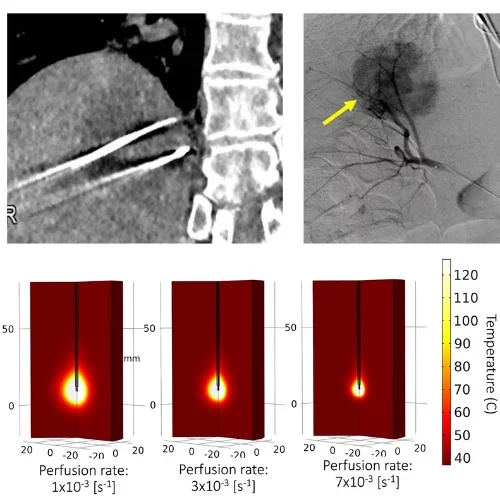
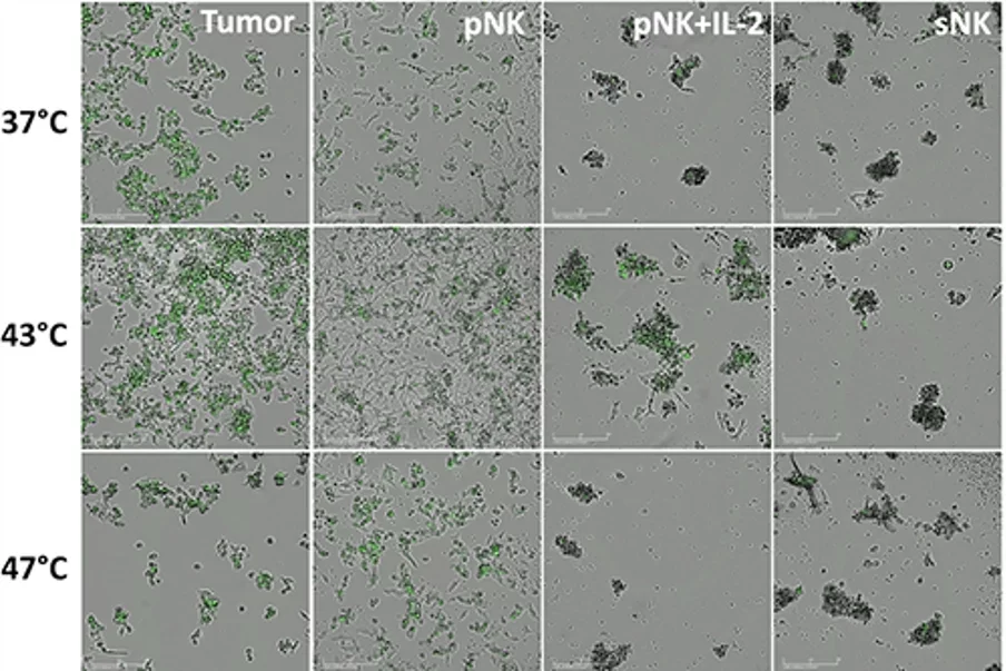
Image-guided immunotherapy to treat early-stage hepatocellular carcinoma (HCC)
Non-resectable early-stage HCC are commonly treated with minimally-invasive, image-guided cancer therapies. While many of these therapies are curative in intent (thermal ablation, radiation segmentectomy) with good long-term outcomes, a subset of HCCs with stem-like features are resistant to these treatment modalities. We are currently studying the potential of using adjuvant NK-cell based immunotherapy to treat these more aggressive HCC cells after traditional locoregional therapy. We work closely with the Tumor Immunology section in the UCLA School of Dentistry.
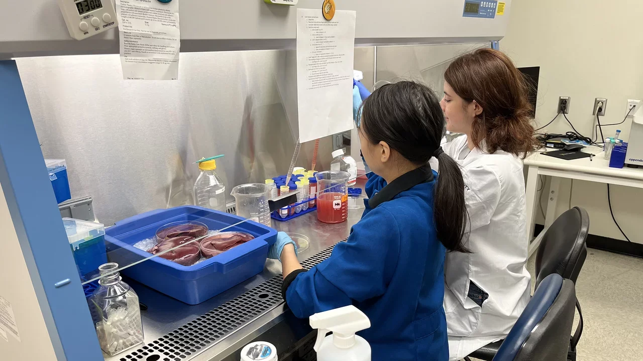
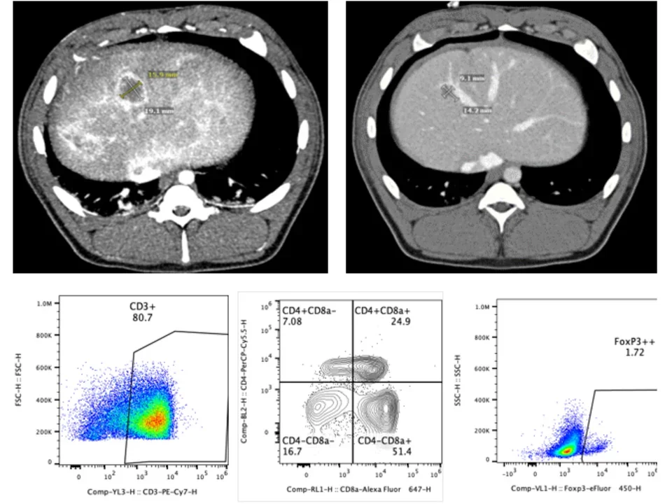
Large animal validation of minimally-invasive cancer therapy
The majority of pre-clinical oncology work has lay within the realm of small animals such as rodents. While they have a role for drug development and validation, rodents are not well equipped to validate the catheters and antennas/probes used for minimally-invasive image-guided cancer therapy. We use large animal platforms to validate the efficacy of locoregional therapy. We collaborate closely with the UCLA Translational Research and Imaging Center (TRIC), which has a dedicated research-only Siemens Prisma 3T MRI, Siemens Somatom Definition CT, and a Siemens Artis Zeego Robotic Arm system for angiography. Protocols are identical to those use clinically to facilitate translation to our own patient population. We also work with the UCLA JCCC Flow Cytometry Core to optimize immune assessment tools for large animals, including a porcine-specific CyTOF panel.
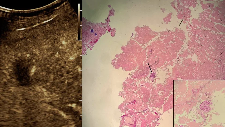
Biomarker analysis to predict responses to locoregional therapy
As interventional radiologists, we have a wide range of minimally-invasive cancer therapies at our disposal. A key goal in our lab is to identify biomarkers that predict response to locoregional therapy, and separate out who is a responder and who is not. We can use cross sectional imaging (CT/MRI), blood samples, and needle-biopsies to help predict who will respond well to locoregional therapies and who will not. We collaborate with multi-omics labs on campus, as well as imaging labs (MRRL) to identify predictive biomarkers for various diseases and treatment modalities.